
Hip Dysplasia in Dogs

 Hip dysplasia is a common joint disease of dogs. It results from abnormal development of the hip joint as a puppy grows. Symptoms include moderate to severe pain and stiffness in one or both hips and can appear at any age. Hip dysplasia is hereditary and is found in many animals (including cats), but it primarily affects large breed dogs such as German shepherds, Labradors, golden retrievers, Rottweilers, great Danes and St. Bernards.
Anatomy of a Dog's Hip Joint
The dog’s hip joint is where the femur (leg bone) attaches to the pelvis (hip bone). It is a ball and socket joint in which the almost spherical top of the femur (the femoral head) fits into the acetabulum, a cup-like depression in the pelvis. A layer of cartilage covers the joint surfaces, allowing smooth, nearly frictionless action over a wide range of motion. A fibrous joint capsule and several strong straps of tissue (ligaments) help keep everything in place. See figure.
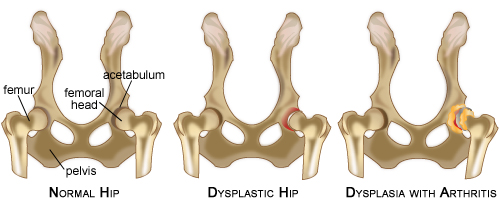
Dogs affected by hip dysplasia are born with normal hips that then develop abnormally during rapid growth. In the dysplastic hip, the ball and socket do not meet properly. The acetabulum is too shallow and the femoral head too flat. The connective tissues supporting the joint may be lax. These abnormalities yield an unstable joint, and degenerative joint disease is the result. Abnormal motion in the joint causes accelerated wear. Cartilage damage occurs, joint tissues are inflamed, and bone spurs form. This causes pain. Degenerative changes tend to increase the instability of the hip, and a vicious cycle ensues.
Signs of Canine Hip Dysplasia
While the occasional dog shows signs in adolescence (i.e., at 5-10 months of age), many dogs live just fine with hip dysplasia until late middle age, when years of wear and tear finally add up. Signs may include:
- A stilted hind-limb gait
- Stiffness or difficulty rising
- Increased weight bearing on the fore-limbs
- A characteristic “hip swivel” gait
- Decreased activity
- A tendency to bunny hop up steps
- Loss of muscle (atrophy) over the hips and thighs
Signs of discomfort may wax and wane, becoming worse after strenuous exercise. As things progress, lameness becomes more persistent. Older dogs tend to gain weight and lose muscle, which accentuates the joint instability.

Diagnosis of Hip Dysplasia in Dogs
Your veterinarian will start with a complete physical examination and medical history. He or she will evaluate your dog’s gait and then perform an orthopedic examination, checking for muscle atrophy, pain, stiffness, or abnormal clicking or popping around the hips. He or she will also want to check for problems in other joints as well as for neurologic conditions that might mimic hip dysplasia.
Hip dysplasia is diagnosed by x-ray. It is important to note that the appearance of a dog’s hips on x-rays is a poor predictor of how much pain a dog has. A dog with ongoing lameness may have only minor changes on x-rays, while a dog with terrible hip films may run, jump, and play as if nothing is wrong. “Dogs don’t walk with their x-rays” is a common saying among veterinarians. Timing of the x-rays can make a difference. Under age 2, a dog’s bones and joints that are still maturing, so standard hip films performed before this point can have unpredictable results. Normal hips at age 2 do not rule out hip problems by age 10.
OFA and PennHIP for Canine Hip Dysplasia
There are currently two standardized paradigms for evaluating hip dysplasia in dogs. The best-known one is administered by the Orthopedic Foundation for Animals (OFA) and a newer one is called PennHIP. Both rely on the fact that hip dysplasia is inherited. Testing dogs for hip dysplasia and allowing only those with good hips to breed has greatly reduced, but not eliminated, the incidence of hip dysplasia in some susceptible breeds. OFA x-rays can be taken by most veterinarians using certain guidelines and then are sent to a certified veterinary radiologist for analysis. PennHIP x-rays require special equipment and training, so they are typically only administered by a veterinary specialist. OFA x-rays are most valid in dogs aged 2 years and above. The PennHIP method is much more sensitive and can be performed on pups as young as 16 weeks of age.
Treatment of Canine Hip Dysplasia
A few simple lifestyle changes are crucial for any dog with degenerative joint disease. Keeping your dog slim minimizes stress on unstable hips. Regular amounts of low-impact exercise alleviate symptoms by decreasing stiffness and strengthening the muscles that support the hip. Warm-up and cool-down periods before and after exercise are essential. Consult your veterinarian for specific recommendations.
Medical options for hip dysplasia are the same as with any degenerative joint disease in which the goal is to relieve symptoms, maintain function, and slow the progression. Click here to learn more about the medical management of degenerative joint disease.
There are 3 surgical treatments for hip dysplasia, and the decision to operate depends on the age of the dog and the severity of the condition. As most are highly specialized and complex surgeries, cost can be a factor as well. Again, your veterinarian can advise you about the options for your dog.
The 3 major surgical treatments are:
- Triple Pelvic Osteotomy (TPO).
- Total Hip Replacement.
- Femoral Head and Neck Excision (FHNE).
The TPO is performed on dogs under 10 months of age with x-rays that show severe hip laxity without any degenerative changes. The procedure, which must be performed by a specialist, involves cutting the pelvis in three places and repositioning the hip socket so it better covers the femoral head. This is a major reconstructive surgery that is expensive but can produce excellent results.
This surgery is for mature dogs with hip dysplasia that have developed severe degenerative joint disease. It is much like hip replacement in humans. The existing hip joint is completely removed and replaced with an artificial joint. It is the only surgery that can produce normal hip function once degenerative changes have set in. This surgery is only performed at surgical specialty centers and is very expensive, but it can have excellent results.
FHNE is a procedure in which the head of the femur is surgically removed. The body eventually forms a fibrous pseudo-joint where the hip was. Range of motion and stability is compromised, but the pain is gone. This procedure can work very well in small- to medium-sized dogs (less than 40-50 pounds) and is a good alternative when the cost of total hip replacement is prohibitive.
Prevention of Hip Dysplasia in Dogs
Screening and selective breeding programs are widely recognized as the best means of preventing hip dysplasia. Parents with radiographically sound hips can still have pups with poor hips but the probability is smaller. When purchasing a purebred dog, it is important to look at the OFA or PennHIP scores going back 3-4 generations.
Certain measures may help decrease the incidence of hip dysplasia in dogs that are genetically susceptible to it. Excess feeding and overly rich diets have been shown to increase hip dysplasia in genetically predisposed puppies. Therefore, large breed puppies aged 2-8 months should be kept slender and given food that is specially formulated for large breeds. Consult your veterinarian for dietary recommendations. It is also thought that repeated strenuous exercise may put undue stress on susceptible hips. While moderate exercise such as running and swimming are excellent for strong muscles and joints, strenuous exercise such as jumping and frisbee-chasing should be avoided in the pup’s first year.
Hip Dysplasia isn’t preventable, but with proper care, most dogs with hip dysplasia can enjoy a good quality of life for many years to come.

You May Also Like These Articles:
Anterior Cruciate Ligament (ACL) Injury in Dogs
Causes of Lameness in Dogs: An Overview
Medial Fragmented Coronoid Process
Osteochondritis Dessicans of the Humeral Condyle (OCD)
Panosteitis in Dogs: Growing Pains
Disclaimer: This website is not intended to replace professional consultation, diagnosis, or treatment by a licensed veterinarian. If you require any veterinary related advice, contact your veterinarian promptly. Information at DogHealth.com is exclusively of a general reference nature. Do not disregard veterinary advice or delay treatment as a result of accessing information at this site. Just Answer is an external service not affiliated with DogHealth.com.
Notice: Ask-a-Vet is an affiliated service for those who wish to speak with a veterinary professional about their pet's specific condition. Initially, a bot will ask questions to determine the general nature of your concern. Then, you will be transferred to a human. There is a charge for the service if you choose to connect to a veterinarian. Ask-a-Vet is not manned by the staff or owners of DogHealth.com, and the advice given should not delay or replace a visit to your veterinarian.